
Ultrasound-guided hip injections are a minimally invasive procedure using medical ultrasound to accurately deliver medications into the hip joint. This technique enhances precision, reduces complications, and provides real-time visualization, making it a safe and effective option for both diagnostic and therapeutic purposes in managing hip-related conditions.
1.1 Definition and Overview
Ultrasound-guided hip injections are a medical procedure where ultrasound technology is used to precisely guide the insertion of a needle into the hip joint or surrounding tissues. This technique allows for real-time visualization of the injection site, ensuring accurate placement of corticosteroids, hyaluronic acid, or other therapeutic agents. Unlike traditional landmark-based injections, which rely on anatomical landmarks and palpation, ultrasound guidance enhances accuracy and minimizes the risk of complications. The procedure is commonly used to diagnose and treat various hip conditions, including osteoarthritis, tendinopathy, and inflammatory arthritis. It is also employed for pain relief and to reduce inflammation in the hip joint or adjacent structures, such as the bursae or tendons.
The use of ultrasound in hip injections has gained popularity due to its ability to improve outcomes and reduce potential side effects. By providing a clear image of the joint and surrounding tissues, ultrasound ensures that the injected medication is delivered directly to the target area. This approach is particularly beneficial for patients with complex hip anatomy or those who have not responded to previous treatments. Overall, ultrasound-guided hip injections represent a safe, effective, and minimally invasive option for managing hip-related pain and inflammation.
1.2 Medical Uses and Applications
Ultrasound-guided hip injections are primarily used to diagnose and treat a variety of hip-related conditions. One of their most common applications is in the management of hip osteoarthritis, where corticosteroids or hyaluronic acid injections are administered to reduce pain and inflammation. They are also effective in addressing hip tendinopathy, particularly in the gluteal or iliotibial band tendons, where precise medication delivery facilitates healing and symptom relief.
Inflammatory arthritis, such as rheumatoid arthritis, benefits from these injections as they allow direct delivery of anti-inflammatory agents into the joint, helping to control synovitis and preserve joint function. Additionally, ultrasound-guided injections are used for diagnostic purposes, such as joint aspiration to analyze synovial fluid for infection or crystal arthropathy.
These injections are also employed in pain management for conditions like trochanteric bursitis, where steroids are injected into the inflamed bursae surrounding the hip. Furthermore, they play a role in postoperative analgesia, such as in lumbar erector spinae plane blocks, to alleviate pain following hip replacement surgeries. Overall, ultrasound-guided hip injections offer a versatile and precise approach to treating hip-related pain and inflammation, enhancing both diagnostic accuracy and therapeutic outcomes.

Procedure Overview
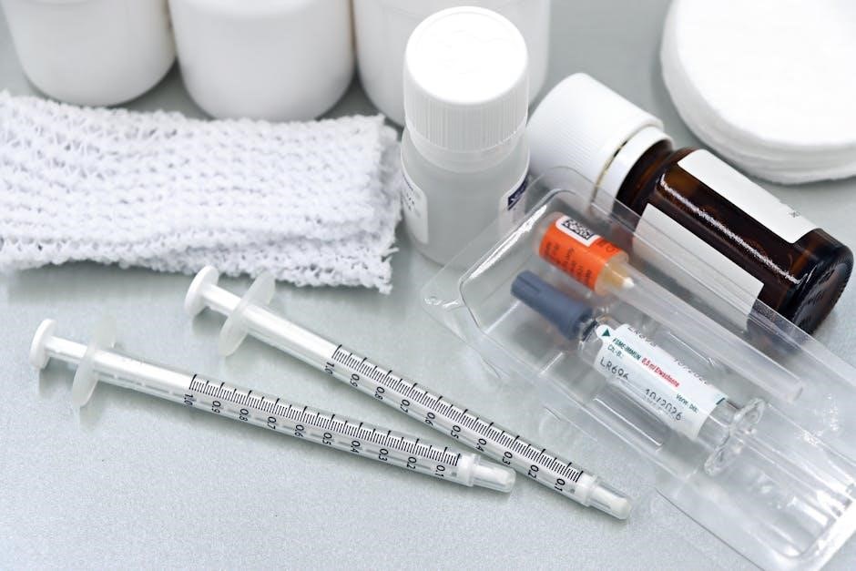
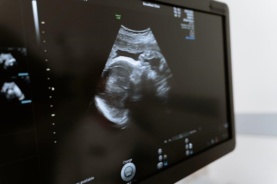
Ultrasound-guided hip injections involve patient preparation, positioning, and ultrasound imaging to visualize the hip joint. A sterile needle is guided under real-time ultrasound to administer medication into the target tissue. Post-procedure monitoring ensures safety and efficacy, minimizing complications and optimizing therapeutic outcomes.
2.1 Step-by-Step Process
The procedure begins with patient preparation, including cleaning and draping the hip area to maintain sterility. The patient is positioned in a supine or lateral decubitus position, depending on the approach. A high-frequency ultrasound transducer is applied to visualize the hip joint anatomy, such as the femoral head, acetabulum, and surrounding soft tissues. The injection site is identified, and local anesthesia is administered to minimize discomfort. Under real-time ultrasound guidance, a sterile needle is inserted through the skin and advanced to the target tissue, such as the intra-articular space or peri-tendinous region. The medication, typically a corticosteroid or hyaluronic acid, is slowly injected while monitoring the spread on the ultrasound screen. Post-injection, the needle is withdrawn, and gentle pressure is applied to the site to prevent bleeding. The patient is monitored for a short period to ensure no immediate complications arise. The entire process is designed to ensure accuracy, safety, and effectiveness in delivering therapeutic agents directly to the affected area.

2.2 Role of Ultrasound in Guiding the Injection
Ultrasound plays a pivotal role in guiding hip injections by providing real-time, high-resolution imaging of the joint and surrounding tissues. This technology allows practitioners to precisely visualize the needle placement, ensuring the medication is delivered directly to the target area, such as the intra-articular space or peri-tendinous regions. By using ultrasound, the practitioner can identify anatomical landmarks, such as the femoral head, acetabulum, and synovial tissue, and avoid vital structures like nerves and blood vessels. The real-time feedback enables adjustments during the procedure, enhancing accuracy and minimizing the risk of complications. Additionally, ultrasound guidance helps confirm the correct placement of the needle before administering the therapeutic agent. This visualization also aids in monitoring the spread of the medication, ensuring it reaches the intended site effectively. The use of ultrasound not only improves the safety and efficacy of the injection but also reduces the reliance on landmarks alone, making the procedure more reliable and consistent. Overall, ultrasound serves as an essential tool in ensuring precise and safe delivery of medications during hip injections.

Advantages of Ultrasound Guidance
Ultrasound guidance enhances accuracy, minimizing complications by avoiding nerves and blood vessels. It provides real-time visualization, ensuring precise medication delivery. This method improves treatment efficacy, reduces procedural pain, and offers a safer alternative to blind injections, making it a preferred choice for hip injections.
3.1 Increased Accuracy and Precision
Ultrasound guidance significantly enhances the accuracy and precision of hip injections by providing real-time visualization of the injection site. This allows healthcare providers to pinpoint the exact location for medication delivery, reducing the risk of misplacement. Unlike landmark-based techniques, which rely on external anatomical references, ultrasound guidance enables direct visualization of soft tissues, tendons, and joint spaces, ensuring that the needle is placed accurately within the target area. Studies have consistently shown that ultrasound-guided injections achieve higher accuracy rates compared to blind or landmark-guided injections, with success rates often exceeding 90%. This precision is particularly important in the hip joint, where the surrounding anatomy is complex and the target area is deep. By using ultrasound, practitioners can avoid nearby nerves and blood vessels, further improving safety. The real-time feedback provided by ultrasound also allows for immediate adjustments during the procedure, ensuring optimal placement of the injected medication. This level of precision not only improves the effectiveness of the treatment but also minimizes the risk of procedural complications, making ultrasound-guided hip injections a preferred method for both diagnostic and therapeutic purposes.
3.2 Reduced Risk of Complications
Ultrasound-guided hip injections significantly reduce the risk of complications compared to traditional landmark-based methods. By providing real-time visualization, ultrasound allows practitioners to avoid critical structures such as nerves, blood vessels, and tendons, minimizing the likelihood of accidental damage. This precise guidance lowers the incidence of adverse events like hematoma, infection, and nerve injury, which are more common in blind injection techniques. Studies have demonstrated that ultrasound-guided injections have a lower complication rate due to improved accuracy and the ability to adjust the needle placement dynamically. Additionally, the ability to visualize the injection site ensures that the medication is delivered directly into the target tissue, reducing the risk of extravasation or misplacement. The enhanced safety profile of ultrasound-guided injections makes them a preferred option for both diagnostic and therapeutic procedures in the hip joint. This method is particularly beneficial in complex anatomical regions like the hip, where surrounding structures are delicate and vulnerable to injury. Overall, the use of ultrasound guidance in hip injections has been shown to enhance patient safety and reduce the potential for complications, making it a cornerstone of modern interventional pain management.
3.3 Real-Time Visualization of the Injection Site
Ultrasound-guided hip injections significantly reduce the risk of complications compared to traditional landmark-based methods. By providing real-time visualization, ultrasound allows practitioners to avoid critical structures such as nerves, blood vessels, and tendons, minimizing the likelihood of accidental damage. This precise guidance lowers the incidence of adverse events like hematoma, infection, and nerve injury, which are more common in blind injection techniques. Studies have demonstrated that ultrasound-guided injections have a lower complication rate due to improved accuracy and the ability to adjust the needle placement dynamically. Additionally, the ability to visualize the injection site ensures that the medication is delivered directly into the target tissue, reducing the risk of extravasation or misplacement. The enhanced safety profile of ultrasound-guided injections makes them a preferred option for both diagnostic and therapeutic procedures in the hip joint. This method is particularly beneficial in complex anatomical regions like the hip, where surrounding structures are delicate and vulnerable to injury. Overall, the use of ultrasound guidance in hip injections has been shown to enhance patient safety and reduce the potential for complications, making it a cornerstone of modern interventional pain management.

Common Approaches in Hip Injections
Ultrasound-guided hip injections commonly use the anterior and lateral approaches. The anterior approach involves supine positioning with an anterior sagittal view, while the lateral approach uses a lateral view with the patient on their side. Both enhance precision and minimize complications, improving therapeutic outcomes.
4.1 Anterior Approach
The anterior approach in ultrasound-guided hip injections is a widely used technique due to its accessibility and clear visualization of the hip joint. Patients are typically positioned in a supine manner, with the hip in a neutral position to facilitate easy access. The ultrasound transducer is placed in the anterior region of the hip, allowing for an anterior sagittal view of the joint. This view provides a detailed image of the femoral head, acetabulum, and the joint capsule, making it easier to guide the needle accurately.
The needle is inserted under real-time ultrasound guidance, ensuring precise placement of the medication into the intra-articular space. The anterior approach is particularly favored for its minimal risk of complications, as it avoids major neurovascular structures. Additionally, this method allows for excellent visualization of the needle tip, reducing the likelihood of procedural errors. The anterior approach is often preferred in clinical practice due to its simplicity and effectiveness in delivering therapeutic agents directly into the hip joint;
Studies have shown that the anterior approach, when performed under ultrasound guidance, enhances the accuracy of injections compared to landmark-based techniques. This method is especially beneficial for patients with hip osteoarthritis, inflammatory arthritis, or tendinopathy, as it ensures targeted delivery of corticosteroids or hyaluronic acid, leading to improved symptom relief and functional outcomes.
4;2 Lateral Approach
The lateral approach in ultrasound-guided hip injections is another commonly utilized method, offering a different perspective for needle placement. Patients are typically positioned in a lateral decubitus position, with the affected hip facing upwards. This positioning allows for easy access to the lateral aspect of the hip joint.
The ultrasound transducer is placed along the lateral thigh, providing a clear view of the femoral head, acetabulum, and the surrounding soft tissues. The needle is inserted under real-time ultrasound guidance, ensuring precise delivery of the therapeutic agent into the intra-articular space. The lateral approach is advantageous as it minimizes the risk of damaging nearby neurovascular structures, such as the femoral artery and vein, which are located more medially.
One of the key benefits of the lateral approach is its ability to provide excellent visualization of the joint space, particularly in patients with severe hip deformities or obesity, where the anterior approach may be less effective. Additionally, this method allows for stable positioning of the needle, reducing the likelihood of procedural complications. The lateral approach is often preferred in clinical settings where patient anatomy or condition necessitates an alternative to the anterior technique, ensuring effective and safe delivery of medications for various hip-related conditions.
Clinical Efficacy in Various Conditions
Ultrasound-guided hip injections demonstrate strong clinical efficacy in treating hip osteoarthritis, tendinopathy, and inflammatory arthritis. They provide targeted delivery of therapeutic agents, reducing pain and inflammation while improving joint function. This minimally invasive approach offers significant benefits for patients with chronic hip conditions, enhancing quality of life and mobility.
5.1 Efficacy in Treating Hip Osteoarthritis
Hip osteoarthritis is a common degenerative condition characterized by cartilage breakdown, pain, and reduced mobility. Ultrasound-guided hip injections have emerged as an effective treatment option, offering targeted delivery of therapeutic agents directly into the joint. Studies demonstrate that these injections, particularly those involving corticosteroids or hyaluronic acid, significantly reduce inflammation and improve joint lubrication, thereby alleviating pain and stiffness.
Clinical evidence highlights the ability of ultrasound-guided injections to provide long-term symptom relief in patients with moderate to severe hip osteoarthritis. For instance, intra-articular injections of hyaluronic acid derivatives, such as hylan G-F 20 (Synvisc), have shown promise in delaying disease progression and enhancing functional recovery. The precision of ultrasound guidance ensures that the medication is deposited accurately, maximizing therapeutic outcomes while minimizing systemic side effects.
Research, including a study by DD Cherkes-Zade in 2010, supports the efficacy of ultrasound-guided injections in improving quality of life for patients with hip osteoarthritis. These procedures are particularly beneficial for individuals who do not respond adequately to oral medications or lifestyle modifications; By combining real-time visualization with advanced injection techniques, ultrasound-guided hip injections offer a safe and reliable solution for managing the symptoms of hip osteoarthritis.
5.2 Efficacy in Managing Hip Tendinopathy
Ultrasound-guided hip injections have demonstrated significant efficacy in managing hip tendinopathy, a condition characterized by inflammation and degeneration of the tendons surrounding the hip joint. This minimally invasive approach allows for precise delivery of therapeutic agents, such as corticosteroids or platelet-rich plasma (PRP), directly to the affected tendons, ensuring optimal results.
Studies suggest that ultrasound guidance enhances the accuracy of injections, reducing the risk of misplacement and improving clinical outcomes; For instance, the Nashville Sound technique, developed for ultrasound-guided hip injections, emphasizes minimal learning curves and high success rates. Real-time visualization enables practitioners to target specific tendon regions, such as the gluteus medius or iliotibial band, which are commonly affected in hip tendinopathy.
Clinical evidence, including a study by Julie M. Labrosse, highlights the effectiveness of ultrasound-guided injections in reducing pain and improving functional mobility in patients with hip tendinopathy. The use of corticosteroids helps alleviate inflammation, while PRP therapy promotes tendon healing by stimulating collagen synthesis and tissue repair.
Overall, ultrasound-guided hip injections offer a safe and efficient treatment option for hip tendinopathy, providing both immediate and long-term relief from symptoms. This approach is particularly beneficial for patients who do not respond adequately to conservative therapies or who require targeted intervention to restore normal hip function.
5.3 Efficacy in Reducing Inflammatory Arthritis Symptoms
Ultrasound-guided hip injections have proven to be highly effective in reducing symptoms associated with inflammatory arthritis, such as rheumatoid arthritis and psoriatic arthritis. By enabling precise delivery of anti-inflammatory medications directly into the hip joint, this technique minimizes systemic side effects and maximizes therapeutic benefits.
Corticosteroids and biologic agents are commonly administered via ultrasound guidance to target inflamed synovial tissue, a hallmark of inflammatory arthritis. Real-time visualization ensures accurate placement of the needle, reducing the risk of injection into surrounding soft tissues. This precision is particularly critical in cases where joint inflammation may obscure anatomical landmarks.
Clinical studies demonstrate that ultrasound-guided injections lead to significant improvements in pain reduction, joint mobility, and quality of life for patients with inflammatory arthritis. For instance, a study comparing ultrasound-guided injections to traditional landmark-based methods found greater symptom relief and faster recovery times in the ultrasound-guided group.
Furthermore, the ability to visualize the injection site in real time allows practitioners to assess the extent of inflammation and adjust treatment accordingly. This personalized approach enhances the efficacy of the procedure and ensures optimal outcomes for patients struggling with chronic inflammatory conditions.
Overall, ultrasound-guided hip injections represent a valuable treatment option for managing inflammatory arthritis, offering both immediate symptom relief and long-term benefits for joint health.